- Industries
Industries
- Functions
Functions
- Insights
Insights
- Careers
Careers
- About Us


Dual Energy Imaging


What is Dual Energy Imaging?
Recent developments in flat-panel detector technology have reignited interest in cutting-edge uses, including dual-energy imaging. Dual-energy imaging allows for the selective imaging of soft tissue and bone tissue, two clinically essential materials. Energy spectra discrepancies for capturing independent images are used to determine energy-dependent differences between two materials to their atomic level. Dual-energy imaging is a technique that creates a single image by combining two X-ray photons with different energies. Due to their various energy absorption characteristics, which enables the distinction between materials and tissues. The two energy levels enter the object or the human body at multiple depths,resulting in images with varying information that can be merged to create a composite image. This information can be utilized to distinguish between tissues or materials similar to one another, such as bone and metal, as well as to emphasize the presence of particular conditions, such as tumors, foreign objects or inflamed areas. Dual-energy imaging can benefit from numerous medical specialties, including radiology, cardiology, orthopedics, cancer, gastrointestinal, and nephrology. Not only for medical purposes but also Dual Energy imaging techniques are being used in different sectors, such as industrial inspection, security screening, art conservation, geological survey, food quality control, etc.
Dual Energy Imaging Techniques
The technical process of Dual Energy Imaging involves the following steps:X-ray Source: Two X-ray sources with different energy spectrums are used to acquire images.
Data Acquisition: Data is obtained from X-ray sources, creating two separate datasets and then processing.
Image Reconstruction: The data from the two X-ray sources are processed and reconstructed into individual images representing different energy spectrums.
Image Fusion: The two images are combined and fused to produce a single, more detailed and accurate picture.
Image Analysis: The final image is analyzed to determine the composition and density of the tissues and materials present. The technical process of DEI utilizes sophisticated algorithms and software to combine and analyze the data from the two Xray sources, producing more accurate and detailed images. The technique can help diagnose and treat various medical and non-medical conditions.
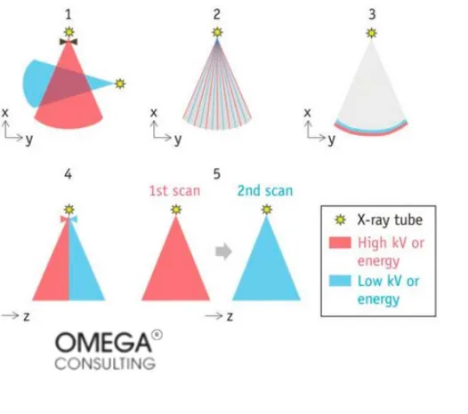
Exhibit 1: Five different methods of data acquisition
X-ray tube could be divided into high kv of energy and low kv of energy. The high-energy end of the spectrum is determined by the KV (kilovoltage) applied to the x-ray tube.
About Dual Energy CT
A. What is the Dual Energy CT?Dual-energy computed tomography (CT), or spectral CT, is a type of dual-energy imaging that uses X-rays to produce detailed cross-sectional images of the body. Whereas conventional single-energy CT has a single image set, dual-energy data (attenuation values at two energy spectra) can be used to reconstruct numerous image types : Images with weighted averages (simulating single energy spectra), virtual images with single energy (attenuation at a single photon energy rather than a range). Photos of material deterioration (mapping or removing substances of known attenuation characteristics, such as iodine, calcium, or uric acid). Substituted non-contrast image (iodine removed).
The amount of iodine (iodine maps) Calcium suppression (removing calcium); Uric acid suppression (removing uric acid); Calcium suppression (removing calcium); Uric acid suppression (removing uric acid); Sufficient Zeff maps for atomic numbers.
Dual Energy CT Techniques
The Dual Energy Computed Tomography (DECT) process involves the following techniques:Material Decomposition: The data from the two X-ray sources is processed to separate the contribution of different materials in the images, such as bone, soft tissue, and contrast agents.
Image Registration: The two images acquired from the different energy spectrums are aligned and registered to produce a combined image.
Image Subtraction: The images from the two energy spectrums can be subtracted to highlight specific structures or materials.
Material Mapping: The composition and density of the tissues and materials in the image can be mapped and displayed in different colors or transparency levels.
Spectral Unmixing: The data from the two energy spectrums can be used to determine the relative proportions of different materials in the image, such as the amount of calcium in bones.
Exhibit 3: Single-Energy Image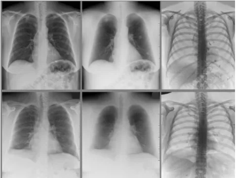
On the left is the conventional single energy image; in the middle is the bone subtracted “soft-tissue only” image; on the right is the soft-tissue subtracted “boneonly” image
Barriers to Dual Energy CT:1. Cost of hardware and software upgrades
2. Limited availability of dual energy CT machines3. Difficulty incorporating dual energy imaging into clinical workflow
4. Need for specialized training for radiologists and technologists
5. Variability in image quality and diagnostic accuracy
6. Higher radiation dose compared to conventional CT scans
7. Difficulty in interpreting and integrating dual-energy images with other imaging modalities.
A survey was circulated to ten radiology departments and shared on two national professional cooperation mailing lists. It examined how DECT is currently used in adult and pediatric settings, along with any usage obstacles and recommendations for solutions. The survey results are shown below.
Exhibit 4: Options for Encouraging DECT
In the graph below, usage in adult and pediatric settings, and reflecting on that people nowadays have a better understanding of the benefits for DECT operations. And encouraging DECT in the education progress, but less encouraging DECT in helping medical experts.
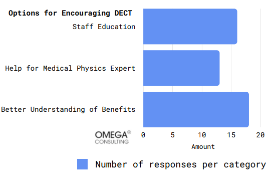
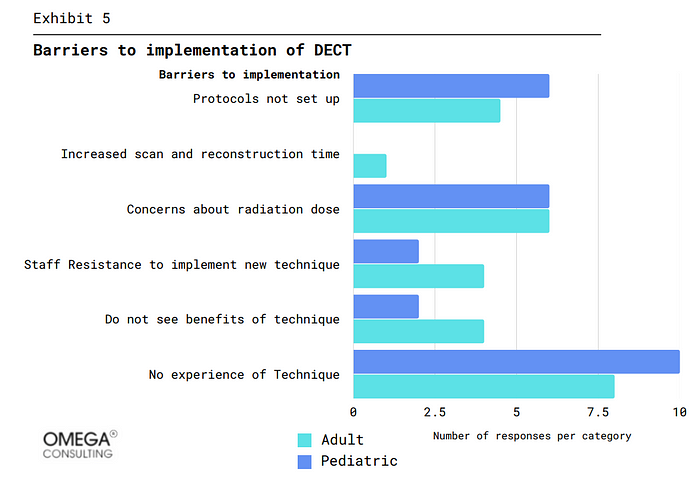
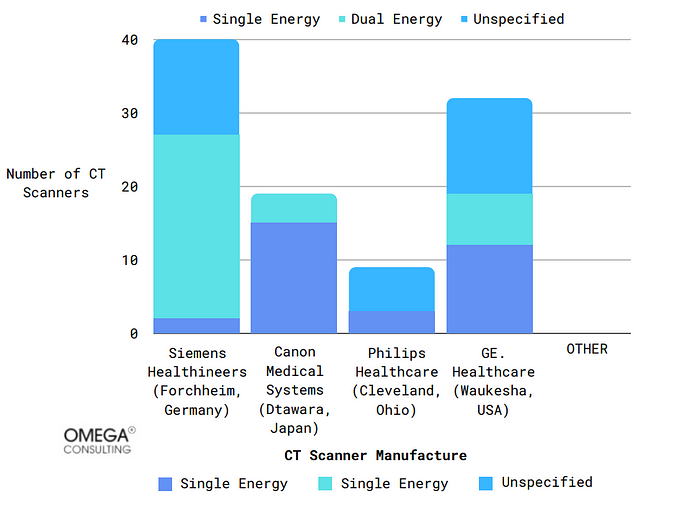
Exhibit 6: CT Scanners split by manufacturer and DECT capability
CT Scanner Manufacture Number of CT Scanners Siemens Healthineers (Forchheim, Germany) Canon Medical Systems (Dtawara, Japan) Philips Healthcare (Cleveland, Ohio) GE. Healthcare (Waukesha, USA) The market for CT scanners is also reflected in the survey. Exhibit 6 shows the number of different types CT Scanner used. Most firms are using the single-energy CT. Even in Germany and the USA, the use of DECT is not as high as single energy CT. However, it can still be assumed that DECT has a promising future in the medical market and will gradually replace the use of single energy CT.
Application of Dual Energy CT:
Dual Energy imaging can provide important information that cannot be obtained from a single-energy X-ray exam, helping to improve patient care and outcomes.
Radiology: It helps differentiate between soft tissue and bone and can aid in detecting certain types of tumors and foreign objects.
Cardiology can also detect and characterize cardiovascular diseases like coronary artery disease. Using two different energy levels of radiation, the image can distinguish between different tissue types, such as healthy and diseased blood vessels, making identifying and diagnosing cardiovascular diseases easier.
Orthopedics: One of the main applications of dual-energy imaging is detecting and characterizing bone fractures. Using two different radiation energy levels, the image can distinguish between different tissue types, such as bone and soft tissue, making it easier to identify fractures.
Oncology: Another application of dual-energy imaging is detecting and characterizing tumors. Using two different energy levels of radiation, the image can distinguish between different tissue types, such as malignant and benign tumors, making identifying and diagnosing cancer easier.
Gastrointestinal: It can assess the gastrointestinal tract for bleeding, inflammation, or tumors.
Nephrology: It can be used to evaluate the kidneys, ureters, and bladder for stones, tumors, and other abnormalities. Overall, dual-energy imaging is a powerful tool for medical imaging that can provide more detailed and accurate images of the body, helping to improve the diagnosis and treatment of various diseases and conditions.
Other Use Cases of Dual Energy Imaging
A. Industrial Inspection: Dual-energy imaging is also used in industrial inspection for non-destructive testing (NDT) to determine the composition and properties of materials. In this context, dual-energy imaging can differentiate between different types of materials and detect defects such as cracks, voids, or inclusions. The technique is commonly used in the aerospace, automotive, and petrochemical industries to inspect components and structures for signs of corrosion, wear, or damage.B. Dual Energy Imaging in the Agriculture sector: Dual-energy imaging in agriculture has the potential to offer helpful information for maximizing crop yields, enhancing soil management, and raising operational efficiency . It is not widely used in the agriculture sector, but it has the potential to be applied in certain areas. For example, it could be used in soil analysis to differentiate different soil types and determine soil properties such as moisture content, nutrient levels, and pH. Additionally, dual-energy imaging could be used to evaluate the composition of plants and crops and to detect the presence of disease, pests, or other abnormalities.
C. Food Quality Control: Dual-energy imaging can also be used in food quality control to determine food products composition and detect contaminants or other foreign materials. The technique can differentiate between different types of food, such as meat, vegetables, and grains, based on their unique energy absorption properties. This information can be used by food manufacturers, processors, and retailers to ensure that food products meet quality standards, improve production processes, and reduce waste.
D. Security Screening: Dual-energy imaging can be used in security screening, such as airport security or border control, detect weapons, drugs, and other contraband items due to their ability to estimate Effective Atomic Numbers (Zeff), which traditional devices cannot do because they use density-based segmentation, dual-energy X-ray devices are chosen over conventional ones.
E. Geographical surveys: Identifying minerals, rocks, and other subsurface elements and geological mapping formations, are all possible with dual energy imaging in geological surveys.
F. Art Conservation: In art conservation, dual-energy imaging can determine artwork authenticity, examine the underlying layers in a painting, and analyze pigments and materials used.
Advantages of Dual Energy Imaging
Some of the main advantages of dual-energy imaging includes the following:Improved tissue differentiation: Dual-energy imaging allows for better separation of different tissues in the body, making it easier for medical professionals to diagnose conditions.
Reduced radiation exposure: Dual-energy imaging reduces the amount of radiation exposure is required compared to traditional single-energy imaging techniques.
Increased accuracy: Using two different energy levels in dual energy imaging allows for more accurate images and helps reduce the effects of artifacts and noise.
Improved detection of particular conditions: Dual-energy imaging is beneficial in detecting certain conditions, such as bone fractures, metal implants, and certain types of tumors.
Versatility: Dual-energy imaging can be used for various applications, including CT scans, X-rays, and bone densitometry.
Disadvantages of Dual Energy Imaging
Some of the disadvantages of Dual energy imaging:Increased radiation dose: Dual energy imaging requires two separate X-ray exposures, which increases the patient’s overall radiation dose.
Cost: Dual-energy imaging is more expensive than traditional single-energy imaging, requiring specialized equipment and software.
Complexity: The dual energy imaging process is more complex than conventional imaging, requiring specialized training and expertise.
Image artifacts: Dual-energy imaging can produce image artifacts such as noise or distortion, making it difficult to interpret the images accurately.
Limited imaging options: Dual energy imaging is typically only used for certain types of imaging, such as bone density or kidney stone detection, and is probably not appropriate for all kinds of medical conditions .
Time-consuming: Dual-energy imaging takes longer than traditional imaging, which can be inconvenient for patients and may delay the diagnosis or treatment of their condition.
Conclusion
Dual-energy imaging is a rapidly growing field with many applications in the medical, industrial, and scientific sectors. This technique provides valuable information about the composition of materials and tissues, which can be used to improve diagnosis and treatment in the medical field to analyze materials in industrial settings, and to study the Earth and cultural heritage in the scientific field. The prospects for dual energy imaging is positive as technological advances continue to increase the accuracy and reliability of these techniques, and new applications continue to emerge. Integrating dual-energy imaging with other imaging modalities are also expected to lead to the development of even more powerful diagnostic tools. Overall, dual-energy imaging is a valuable tool that has the potential to revolutionize a wide range of fields, and its impact will likely continue to grow in the future.Subscribe
Select topics and stay current with our latest insights
- Functions